
Retinal Diseases
Retinopathy Of Prematurity
The retina is a neural light sensitive layer stretched along the inner side of the back of the eye. We are able to visualize what we see because of the light sensitive receptors embedded in this neural retina. The light falling on the receptors stimulates the receptors, which then generate electrical signals, which are then carried along the neurons(electrical signal carrying nerve cells) in the retina and the optic nerve all the way to the back of the brain to an area known as the occipital lobe where the electrical signals are interpreted and the brain recognizes the form and the color of the generated image.
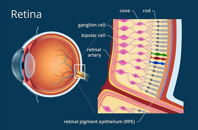
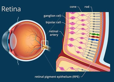
RETINA
What is Diabetic Retinopathy?
Diabetic retinopathy is an eye condition caused by long-standing diabetes that damages the small blood vessels in the retina — the light-sensitive layer at the back of the eye. Over time, this damage can lead to vision loss and even blindness if not detected and treated early.


Why does it occur?
When blood sugar levels stay high for long periods, the tiny blood vessels in the retina become weak and leaky.
Causes For Diabetic Retinopathy:
Swelling of the retina (macular edema)
Poor blood supply to certain areas of the retina
Growth of abnormal new blood vessels that can bleed or scar
Types of Diabetic Retinopathy:
1. Non-Proliferative Diabetic Retinopathy (NPDR) – Early stage, with small leaks or blockages in retinal vessels.
2. Proliferative Diabetic Retinopathy (PDR) – Advanced stage, where abnormal blood vessels grow and can cause bleeding, scarring, or retinal detachment.
Symptoms to watch for:
Early stage: Often no symptoms; only detected in routine eye exams.
Progression: Gradual blurring, floaters, or sudden vision loss, difficulty in distinguishing colour, difficulty in seeing at night.
Treatment Options:
Treatment depends on the stage and severity:
1. Control of diabetes – Strict blood sugar, blood pressure, and cholesterol control is essential at all stages.
2. Laser treatment (Photocoagulation) – Seals leaking vessels and prevents abnormal vessel growth.
3. Injections (Anti-VEGF or Steroids) – Reduce swelling and stop abnormal vessel growth.
4. Vitrectomy surgery – Removes blood, scar tissue, or repairs retinal detachment in advanced cases.
Diagnosis for Diabetic Retinopathy
1. Visual Acuity Testing
A standard eye test to measure clarity of vision.
2. Tonometry
Measures intraocular pressure to assess eye health.
3. Pupil Dilation Exam
Drops are used to widen the pupil, allowing the doctor to examine the retina.
4. Optical Coherence Tomography (OCT)
A detailed imaging test that detects retinal swelling and thickness changes
Frequently Asked Questions:
What Is Retinal Detachment?
The retina is attached to the layer immediately below it called as the choroid. Sometimes the retina can peeled away from the underlying choroid. It leads to vision impairment or blindness if left untreated
Causes for Retinal Detachment:
1) A tear or a hole in the retina (Rhegmatogenous retinal detachment) this can happen in people having a high minus spectacle number(myopia), eye injury, due to ageing, or due to a pull on the vitreous base which may happen after cataract or other ocular surgery or after the YAG laser posterior capsulotomy
2) Some scar tissue pulling on the retina from the inside of the eye(tractional retinal detachment). This may happen in diabetic retinopathy, chronic intraocular inflammation or after eye injuries or after previous eye surgery.
3) Fluid collection beneath the retina (exudative retinal detachment) which may happen in people having high blood pressure, pregnant females who develop eclampsia, with orbital or intraocular tumors, other inflammatory conditions of the eye(uveitis), or after trauma.
Systoms to watch for
1) Flashes of light
2) Floaters
3) A curtain falling across the field of vision.
It is often a painless event, but which necessitates emergency treatment. Treatment instituted within few days after hells in regaining of useful vision. This is because the detached retina loses it light capturing sensitivity progressively. As the duration of the detachment increases, the sensitivity may decrease to such a extent that even after anatomically successful surgical reattachment of the retina fails to grant any useful vision to the patient.
Treatment options
Retinal detachment is treated by
1) vitrectomy with silicon oil or gas injection, followed by endolaser.
In this procedure, done under injection local anesthesia, the vitreous is removed using special vitrectomy machines, and the retina id unfolded and attached using gas bubble or silicon oil bubble. The gas bubble is absorbed by the body in a few days time. In case silicon oil bubble is used, another procedure may need to be performed after a few months to remove the silicon bubble.
2) external surgery using silicon tyres and buckles.
In this procedure doen under injection local anesthesia, a silicon patch is sutured onto the sclera overlying the retinal break. In some cases, if there are multiple breaks or large breaks, then a silicon tyre and a buckle is encircled on the eye ball. This can stay inert for a long time and can help in reattachment of the retina.
3) pneumoretinopexy which may be done under injection local anesthesia, in which a gas bubble is injected to repair the retinal detachment, followed by laser PRP
4) only laser barrage is sometimes done under topical anesthesia. In case of small retinal holes. The laser barrage seals off the retinal hole and prevents the retina from detaching further.
Frequently Asked Questions:
BRVO: Don't Let It Dim Your Vision
What Is BRVO?
Branch Retinal Vein Occlusion is a blockage in one of the smaller veins that drain blood from the retina. It’s the most common type of retinal vein occlusion and typically affects one quadrant of the retina..
Why does it occur?
The blockage prevents blood from draining properly, leading to fluid leakage, retinal swelling (macular edema), and sometimes ischemia (poor blood supply)
What you will experience?
Sudden, painless vision loss or blurring in part of the visual field. Floaters may appear due to bleeding into the vitreous gel.
Causes and Risk Factors
BRVO often occurs at arteriovenous crossings, where a hardened artery compresses a vein, leading to turbulent blood flow and clot formation.
Common causes and risk factors include:
Factor Description
Atherosclerosis: Hardening of arteries compresses veins at crossing points
Hypertension: High blood pressure increases vascular stress
Diabetes: Damages blood vessels and promotes clotting
Glaucoma: Elevated intraocular pressure may contribute
Smoking: Accelerates vascular damage
Age > 50 : Vessel elasticity decreases with age
Obesity: Associated with systemic vascular issues
Blood clotting disorders: Especially in younger patients
Course of Disease
The progression of BRVO varies:
Initial Phase: Sudden vision loss due to hemorrhage and edema.
Intermediate Phase: Macular edema may persist or worsen; ischemia can develop.
Late Phase: Neovascularization (growth of abnormal vessels) may occur, risking vitreous hemorrhage or neovascular glaucoma.
Without treatment, some cases stabilize, but others may lead to permanent vision loss
due to scarring or ischemic damage..
💉 Treatment Options
Treatment focuses on managing complications like macular edema and preventing further vision loss:.
1. Anti-VEGF Injections
Drugs: Ranibizumab (Lucentis), Aflibercept (Eylea), Bevacizumab (Avastin)
Mechanism: Reduce vascular leakage and edema
Effectiveness: ~50% of patients show significant visual improvement
Frequency: Regular injections required for sustained benefit
2. Steroid Implants
Drug: Ozurdex (Dexamethasone)
Mechanism: Reduces inflammation and edema
Duration: Lasts 4–6 months; repeated over 2–3 years
Use: For patients unresponsive to anti-VEGF therapy
3. Laser Therapy
Macular Laser: Targets fluid leakage in the macula
Sectoral Panretinal Photocoagulation: Used if neovascularization develops
Purpose: Prevent further bleeding and preserve peripheral vision
4. Observation
Mild cases may improve spontaneously
Regular monitoring is essential to detect worsening or complications